Having an MRI when you dread enclosed spaces often means either giving up on the exam or enduring it with anxiety that’s hard to manage. Open MRI offers a concrete alternative for claustrophobic patients, with a machine that is open on the sides. Feedback from those who have tried this technology reveals a reality that is more nuanced than the initial promise.
How the shape of the magnet affects the patient’s experience
Have you ever seen a photo of a traditional MRI? A long, narrow tube where the entire body disappears. Open MRI works differently: the magnet takes the shape of a C or a horizontal platform. The patient lies between two surfaces, but their sides remain free.
This lateral openness radically changes the perception of space. Some patients describe the sensation of undergoing an exam on a simple table, with an unobstructed view of the room. A report from France 3 Régions shares the testimony of a patient comparing the old closed MRI to “entering a coffin,” while the open MRI allowed him to see outside throughout the exam.
Several people who shared reviews on open MRI for claustrophobes confirm this immediate relief related to the absence of a tunnel. The difference is evident from the setup: not feeling a wall above the face is enough to lower the anxiety level for most affected patients.
Open MRI or wide-bore tunnel: a common confusion in testimonials
A common trap appears in online feedback. Many patients believe they underwent their exam on an open MRI when they were actually in a wide-bore MRI. Both improve comfort, but their design is entirely different.
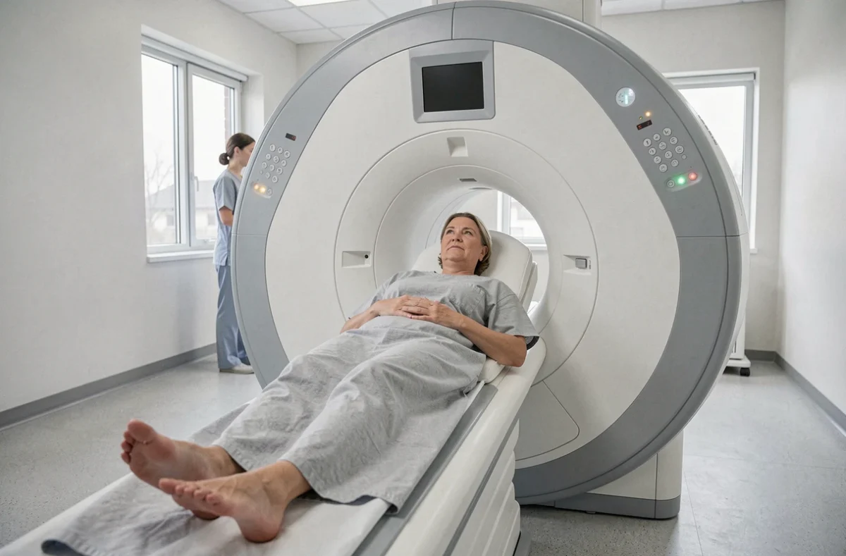
The wide-bore tunnel remains a closed tube, with a more generous diameter and reduced depth compared to older models. The open MRI, on the other hand, eliminates the tube. This distinction matters for people suffering from severe claustrophobia: a wider tunnel may suffice in cases of mild to moderate anxiety, but it does not replicate the sense of space of an open machine.
This confusion complicates the interpretation of testimonials. A patient satisfied with their exam in a wide-bore tunnel does not describe the same experience as a patient who underwent an exam on a C-shaped magnet machine. Before relying on feedback, checking the exact type of machine used helps calibrate expectations.
Why the body region being examined matters as much as the machine
One point that testimonials illustrate well: brain exams remain the most difficult to endure, even on a wide-bore machine. The head must be positioned deep within the machine, which partially negates the gain in space. Literature on wide-bore machines confirms that patients who did not manage to complete their exam were primarily scheduled for a brain MRI.
In contrast, an exam of the knee or shoulder leaves a large part of the body outside the machine. The comfort felt thus varies as much according to the area being explored as according to the type of machine.
Human support and immersion devices: what really makes the difference
The most positive feedback does not only talk about the geometry of the machine. They also mention what happens around: the presence of staff, physical contact, the visual environment.
The report from France 3 Régions describes patients lying on their side, with a view of a calming decor, while a caregiver held their hand. This type of support transforms the exam. Several testimonials converge on a simple observation: the reassuring presence of a caregiver reduces anxiety as much as the openness of the machine.
The medical imaging centers that receive the best feedback generally combine several elements:
- An appropriate positioning of the patient (on their side when possible, feet first rather than head first)
- Maintaining visual or physical contact with the staff during the exam
- Distraction devices: headphones with music, mirror glasses allowing them to see the room, or a viewing screen
These adjustments may seem trivial on paper. In testimonials, they emerge as decisive factors between a completed exam and an interrupted one.

Limitations of open MRI: what patients discover after the exam
Comfort comes at a technical cost. Open machines generally operate at a lower magnetic power than closed high-field MRIs. The direct consequence: the image quality may be lower for certain medical indications.
Patients report having to undergo a follow-up exam in a closed MRI because the images obtained in the open MRI did not provide the necessary resolution for diagnosis. This feedback recurs regularly and generates frustration: the psychological effort made for the initial exam then seems wasted.
The number of open MRIs in France remains limited. Finding a center equipped with one may involve a significant journey, with appointment delays sometimes longer than for a traditional MRI. This scarcity practically limits access to this solution.
When sedation remains an option to consider
For patients whose claustrophobia prevents any exam, even in an open machine, light sedation with an anxiolytic prescribed before the exam is a popular alternative in testimonials. It allows them to undergo the MRI in a standard closed machine, often more effective.
General anesthesia also exists as a last resort, but feedback regarding it is rare. Most patients find a solution before reaching that point, between the choice of machine, the support of staff, and medication preparation.
- Mild claustrophobia: a wide-bore tunnel with appropriate support is often sufficient
- Moderate claustrophobia: open MRI provides significant relief
- Severe claustrophobia: combining open MRI, light sedation, and the presence of a caregiver offers the best success rate according to patient feedback
The choice of machine is not made in isolation. A prior discussion with the radiologist, precisely describing their level of anxiety and the area to be examined, helps guide towards the most suitable configuration. The most serene testimonials almost always come from patients who had prepared this exchange in advance.